Oral paracoccidioidomycosis. A clinical case |
Publicado el: 04/05/2011 13:34:59 |
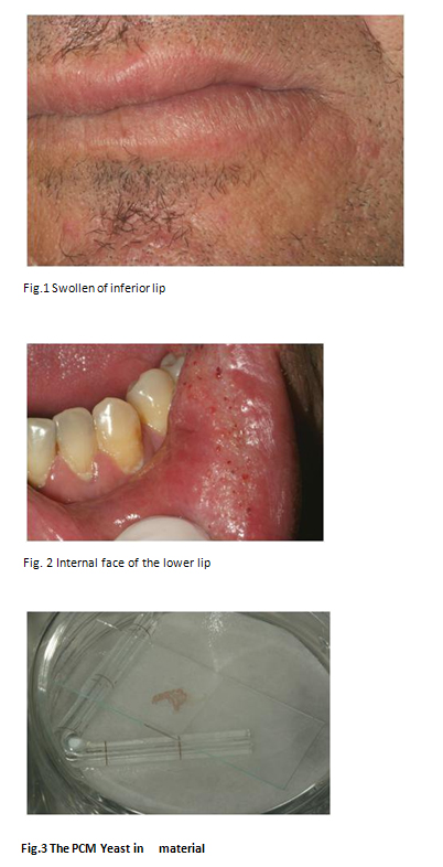
| Article`s Title: ORAL PARACOCCIDIOIDOMYCOSIS. A CLINICAL CASE Running Title:OralParacoccidioidomycosis Key words: paracoccidioidomycosis, oral mucosa, swelling. Authorsname:(1)FILIPE COIMBRA (2)ANA LANHAS LOPES (3)Elaine Massucato (4)Ricardo Faria Almeida (5)Fontes de Carvalho (6)António Felino AcademicPost: (1) Assistant Professor, Faculdade de Medicina Dentária da Universidade do Porto, Portugal. (2) MedicalDentist, Faculdade de Medicina Dentária da Universidade do Porto, Portugal. (3) Assistant Professor, Faculdade de Odontologia de Araraquara, Brasil. (4) Associated Professor, Faculdade de Medicina Dentária da Universidade do Porto, Portugal. (5)Associated Professor, Faculdade de Medicina Dentária da Universidade do Porto, Portugal. (6) Catedratic Professor, Faculdade de Medicina Dentária da Universidade do Porto, Portugal. Mailing address. Rua Nossa Senhora da Hora, 65, 4430-182, Vila Nova de Gaia, Portugal Telefhone Number: +351962492386 e-mail address: filipepacoimbra@gmail.com SUMMARY A middle-aged man complaining of a hard swelling of the oral mucosa of the inferior lip and persistent dry cough was studied by one of us during a visit to theBrazilian Town of Araraquara in the State of S. Paulo. The patient was a laborer with smoking and drinking habits. Long yeast cells with multiple branches were detected at microscopic examination of material scrapped from the mucosal swelling and after further cultures in Sabouraud`s agar. Blood serum double Immunodiffusion and crossimmunoelectrophoresis techniques showed positive reactions against purified antigens extracted from Paracoccidioidesbrasiliensis. Oral administration of Itraconazol led to marked improvement of the oral symptoms. Since this infection can persist for years in human carriers, it is important that the dental practitioner may suspect of its presence in patients comingto Portugal or Spain from South America. Introduction Paracoccidioidomycosis(PCM) is a fungal infection caused by Paracoccidioidesbrasiliensis occurring in Central and South America.In Brazil it predominates in the Southern States namely S. Paulo, Minas Gerais, Rio de Janeiro, Paraná and Rio Grande do Sul(1).The agentlives free on the earthly ground, contaminating humans by inhalation of the spores and giving rise to pulmonary infections that may spread through the lymphatic or bloodroutesto lymph nodes, mouth, skin and adrenal glands(1,2,3,4). Most frequent is the chronic form occurring in male agricultural laborers over thirty-years old, living in bad social conditions associated with malnutrition, alcoholism or over-smoking(5).Clinical symptoms are usually delayed appearing years after contamination when patients have moved to urban centers(2,6). They then often exhibit a dry, persistent cough anddyspnea(3,7), but oral lesions may also occur as the first symptom, located in lips, the oral mucosa, palate and tongue(1,4,8),appearing as swollen mucosal areas sometimes ulcerated. The acute form is present in children or young adolescents of both genders, and If not early detected it may quickly assume serious proportions involving lungs, the tegument, lymph nodes, the lymphatic organs of the digestive tract, spleen, liver and lungs (9,10). Our purpose in the present work is to present a clinical case of chronic PCM. Since cases have been reported abroad in patients having visited South American countries11, it may be useful to know this pathology in Portugal and Spain. CASE REPORT H.Z., a male widowbrick-layer,53 years old, of Caucasian ascent, born in Araraquara (S. Paulo, Brazil), smoker and alcoholic for 30 years, was seen by one of us (Ana Lopes) in the Department of Oral Medicine of the Faculty of Odontology of Araraquara, complaining of a tooth fall one month ago, that was followed by the development of a spongy mass in the tooth cavity. The neighboring inferior lip was swollenand insensitive. (Fig.1). There was a burning sensation in the oral cavity and persistent dry cough. The mucosal surface of the posterior face of the left lower lip was swollen and hardenedwith a reticulated appearancedue to the presence of scattered bleeding points and fissures, extending laterally from the oral commissure for a 7 cm. length to the jugal mucosa (Fig. 2).A firm spherical pediculated mass, 1.5 cm. in diameter, occupied the place of teeth 37 and 38.Very bad oral hygiene with loss of various teeth.Material scrapped from the affected mucosa wasdirectly examined or cultured in Sabouraud's agar. In both cases, long yeast cells with multiple budding were observed under the microscope (Fig.3).In blood serumdouble-immunodifusion (ID) showed positive reaction to an antigenic preparation of P. brasiliensis and negative reactions toward HistoplasmacapsulatumorA. fumigatus. With the crossed-immunoelectrophoresis (CIE) technique results were likewise positive only for P. brasilirensis(1/26 sensitivity). Lung X-rays showed interstitial fibrosis in the form of butterfly wings. The pediculated growth appeared under the microscope as a typical posttraumatic fibroma. Treatmentconsisted of a daily pill of Itraconazol, 200 mg during 15 days, after which there was a marked improvement of the oral lesions. The medication was maintained, and extensive dental care was carried out with extraction of various teeth. Although there was complete regression of local edema, the white color of the mucosa persisted. DISCUSSION This was a typical case of PCMwith involvement of the lung which is usually the primary location in this condition in accordance with the airborne fungal penetration (1,3,4). Lungs are exclusively affected in 25% of the diagnosed patients at the beginning of the infection but are also lesioned in 90% of the multifocal presentations in which the agent has contaminated other organs through blood or lymphatic routes(1,2,7). This probably happened with this patient, a brick-layer for many years working in contact with the soil though being not an agricultural laborer. It may be noted that rareunifocal presentations in the oralregion may occur given the habit of chewing leaves or tooth picking using short branches of plants (12). Direct contamination from person to person or congenitally from parents has never been observe The infection is usually acquired between 10 and 20 years of age, remaining silent until after the patient reaches the third decade. The reactivation is then due to smoking or drinking habits, or a diminished immunity often seen in people living in precarious conditions (7,13). Curiously females are less affected (one woman for 10-25 males) which has been explained by the occurrence of estrogen membrane receptors in the parasite. When activated such receptors do prevent the conversion of the hyphalform occurring in soil into the pathogenic yeast form within body tissues (8).In this present clinical case the lesion of the lip mucosa was, as usual, of the hypertrophic type but devoid of ulceration as sometimes occurs (4). If left untreated the inflammatory process may invade the rest of the mouth inner lining. Palatal lesions have the danger of propagation to the underlying palatal bone(12). As here shown, examination of scrapped material was enough to detect the fungus(15), so that biopsy is usually superfluous. When the latter is performed, histological examination of Grocott-Gomorimethenamine-silver preparations reveals the presence of large black stained yeasts within the cytoplasm of multinucleated giant cells surrounded by multiple spores (4).Serological methods are also mandatory since both ID and CIE have a sensitivity which varies between 85 and 100 % in the pre-treatment stage. In addition, antibody titration of blood serum correlates withthe seriousness of the illness, although its predictive value diminishes a little in the follow-up during treatment (4). As to the medication there are three drug options, amphotericine B (a polyene anti-fungal agent), sulfadiazine (a sulfonamide derivative) and the azolic compounds (14). The latter are the most effective in mild-to-moderate cases with preference for Itraconazol (14), which in this case had a prompt ameliorative action. It should be taken for at least three months and the patient must beseen every six months thereafter, with serological control to insure the virtual eradication of the mycosis.But it should be noted that total cure is problematic given the tendency of the yeast to remain dormant in the body. Therefore follow-up is recommended for some years ahead. In a non-endemic region like Portugal or Spain, the here described clinical symptoms should lead the dental practioner to suspect of other pathologies such as lung tuberculosis or sarcoidosis in which cough and X-ray lesions may coincide with oral alterations (6). On the other hand the occurrence of a persistent lip enlargement is frequent in orofacialgranulomatosis (6). In all these cases, local biopsy is necessary. If however the patient came from a country in which fungal infections are endemic PMC should be taken into consideration. REFERENCES 1.Martins GB, Salum FG, Figueiredo MAS, Cherubini K, Yurgel LS. Paracoccidioidomicose bucal Relato de 3 casos.BraPatol oral 2003 Jul-Set;2(3):22-28. 2. Shikanai-Yasuda MA, Telles filho FQ, Mendes RP, Colombo AL, Moretti ML. Guidelinesinparacoccidioidomicose. RevSocBrasMedTrop. 2006 May-Jun;39(3).297-310. 3.Oliveira Mo, Pistóia AD, Neuhaus C, Veiga Fb. A paracoccidioidomicose na odontologia. Relato de um caso. Saúde 2005; 31 (1-2):10-15. 4. Silvio AM. Paracoccidioidomicose: Atualização epidemiológica, clínica e terapêutica.BrasDermatol78(2):134-150, Mar-Abr.2003. 5. PaniagoAm, Aguiar JI,Aguiar ES, Cunha RV,Pereira GR, Londero AT, Wank B. Paracoccidioidomicose:a clinicalandepidemiologicalstudyof 422 casesobservedin Mato Grosso do Sul.SocBrasMedTrop. 2003 Jul-Aug;36(4).455-9. 6.Palmeiro M, Cherubini K, Yurgel LS. Paracoccidioidomicose-LiteratureReview. Scientia Medica. 2005 Out-Dez;15(4):274-8. 7.Pedroso VS,Vilela C, Pedroso ER, Teixeira AL Paracoccidioidomicosiscompromisingthe central nervoussystem: a reviewoftheliterature. SocBrasMedTrop. 2009 Dec,42(6):692-7. 8.Vieira EMM, Borsatto-Galera B.Manifestações clínicas bucais da paracoccidioidomicose. PatolTrop 2006Jan-Abr;35(1):23-30. 9,Meneses-Garcia A, Mosqueda-Taylor A, Moralesdela Luz R, Rivera LM.Paracoccidioidomicose: reportof 2 cases mimicking scamous cell carcinoma. Oral Surg Oral Pathol Oral RadiolEndod.2002 Nov;94(5):609-13. 10.Ribeiro LC, Hahn RC, Favalessa OC, Tadano T, Fontes CJ. Systemic mycosis:factors associated with death among patients infected with the immunodeficiency virus, Cuiabá, State of MatoGrosso, Brazil, 2005-2008. Rev Soc Bras Med Trop. 2009 Dec;42(6):698-705. 11.Bousquet A, DussartC,Drouillar I, Charbel EC, Boiron P. Imported mycosis. A review of paracoccidioidomycosis. Med Mal infect. 2007 Dec; 37 Suppl 3:S210-4. 12. Castro LGM, Muller AP, Migliari DA. Hard palate perforation: anunusual finding in paracoccidioidomycosis. Int J dermatol 2001:40(4):281:83. 13, Bisinelli JC, Telles FQ, Sobrinho JA, Rapoport A. StomatologicalManifestationsofParacoccidioidomycosis.Bras Otorrinolaringol.2001 Sep;67(6):683-7. 14. Bicalho RN, Santo MF, Aguiar MC, Santos VR. Oral Paracoccidioidomycosis: a retrospective study of 62 Brazilian patients. Oral Dis. 2001 Jan;7(1):56-60. 15.Talhari C, de Sousa JV, Parreira VJ, Reinel D. Talhri S. Oral exfoliativecytology as a rapiddiagnostictool for Paracoccidioidomycosis. Mycoses. 2008 Mar;51(2):177-8.
|
Publicado el: 04/05/2011 13:34:59 |